The Integrated Imaging Facility (NDIIF) at the University of Notre Dame is currently installing two new research instruments with wide-ranging applications in elemental research, materials science, and cancer research. The Light Sheet Fluorescence Microscope (Bruker MuVi SPIM) will be an integral part of the Optical Microscopy Core, while the Transmission Electron Microscope (Thermo Scientific Spectra 300) will help to strategically expand the Electron Microscopy Core.
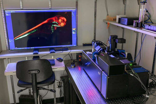
The new Light Sheet Fluorescence Microscope uses innovative photon illumination techniques that can accommodate a variety of samples. This instrument produces three-dimensional images at faster speeds with improved axial resolution over that of traditional confocal microscopes. Additionally, the multimodal microscope can image live samples at high speeds and at high resolution while minimizing phototoxicity and photobleaching. This tool expands the range of research able to be conducted at Notre Dame and assists users in the investigation of cell behaviors, engineered vascularized tissue models, and biorobots.
The new Transmission Electron Microscope provides extremely high resolution scanning transmission electron microscope imaging performance. It is able to resolve the smallest of samples, even down to half of an atom. By utilizing accelerated photons, the Spectra 300 performs rapid diagnostics from several angles to create a three-dimensional projection of the sample at high resolution. The microscope’s imaging quality provides great potential for materials science and semiconductor applications.
“We are ecstatic to have two new state-of-the-art microscopes available on campus. They will help to cement Notre Dame as a major hub for multidisciplinary imaging research in Indiana”, said Bradley Smith, director of the Notre Dame Integrated Imaging Facility and Emil T. Hofman Professor of Science in the Department of Chemistry. “These additions to NDIIF will advance ongoing projects and amplify Notre Dame’s contributions in innovative science research, education, and outreach.”
Funds to purchase the Light Sheet Fluorescence Microscope were awarded to the University by a National Science Foundation standard grant. Jeremiah Zartman, associate professor of chemical and biomolecular engineering, served as principal investigator of the proposal. Additional funds were provided by Notre Dame Research. Funds to purchase the new Transmission Electron Microscope were provided by Notre Dame, with additional diagnostic equipment purchased by NDIIF.
A memorial plaque will accompany the new Transmission Electron Microscope to recognize the significant contributions to the Notre Dame Transmission Electron Microscopy Program made by Sergei Rouvimov, associate research professor of electrical engineering. Rouvimov was the Transmission Electron Microscopy Program Director until his death in May 2020.
The new Light Sheet Fluorescence Microscope is now available to Notre Dame researchers and the new Transmission Electron Microscope will be available for use in October 2021. To learn more, please visit imaging.nd.edu.
Contact:
Joanne Fahey, Director of Research Communications
fahey.17@nd.edu / 574.631.9762
research.nd.edu / @UNDResearch
About Notre Dame Research:
The University of Notre Dame is a private research and teaching university inspired by its Catholic mission. Located in South Bend, Indiana, its researchers are advancing human understanding through research, scholarship, education, and creative endeavor in order to be a repository for knowledge and a powerful means for doing good in the world. For more information, please see research.nd.edu or @UNDResearch.
Originally published by at imaging.nd.edu on August 02, 2021.